LABORATORY STAFF
Suren Seryozhayi Mamyan – Head of the Laboratory, Candidate of Chemical Sciences
Email: suren.mamyan@gmail.com
Garik Georgii Martirosyan – Senior Researcher, Candidate of Chemical Sciences
Email: ggmartirosyan@gmail.com
Manuk Nesteri Nersisyan – Researcher, Candidate of Physical and Mathematical Sciences
Email: manuk_nersisyan@yahoo.com
Astghik Artaki Hovhannisyan – Researcher, Candidate of Chemical Sciences
Email: ahovhan@gmail.com
Gohar Shaheni Hovhannisyan – Researcher
Email: l_oganesyan@mail.ru
Nelli Avagyan – Junior Researcher, Candidate of Technical Sciences
Email: nelli.avagyan80@gmail.com
Arsen Seryozhayi Azizyan – Junior Researcher, Candidate of Chemical Sciences
Email: asazizyan@gmail.com
Syuzi Manveli Grigoryan – Junior Researcher
Email: s.grigoryan0708@gmail.com
Lusine Samveli Harutyunyan – Junior Researcher
Email: lusine.harutyunyan@ro.ru
Arbak Hayki Hovhannisyan – Laboratory Assistant
Email: arbakhovhannisyan@gmail.com
Hayk Arami Aghekyan – Laboratory Assistant
Email: hayk16aghekyan@gmail.com
Gurgen Zhorayi Petrosyan – Laboratory Assistant
Email: gurgen299792458@gmail.com
History of the laboratory
Kurtikyan, the laboratory has studied and continues to study the interaction of heme model systems with various signaling molecules: O2, NO, CO, H2S. Nitrosyl, nitrite, and nitrate complexes of a number of metalloporphyrins with various trans ligands have been discovered and studied. Of great interest are the formation of metal-ligand bonds and the study of the further reactivity of coordinated ligands The use of low temperatures makes it possible to study fast reactions, record intermediate compounds, and obtain their spectral characteristics. To solve problems in organic, inorganic, coordination and polymer chemistry, the laboratory has developed methods that allow for spectral research of solid, liquid and gaseous substances in a wide temperature range (10K -350K). Experiments are performed in the low temperature range using a closed-cycle helium (starting at 10K) refrigeration system and liquid nitrogen (starting at 77K) cryostats. The laboratory has also developed special methods for measuring the IR, UV-Vis and Raman spectra of sublimated films.
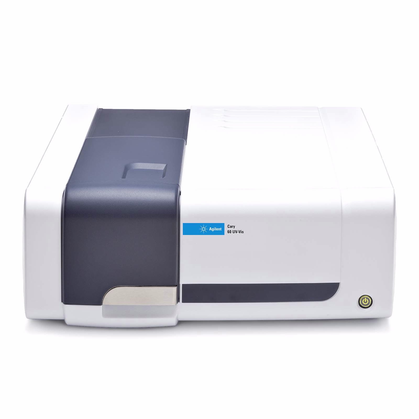
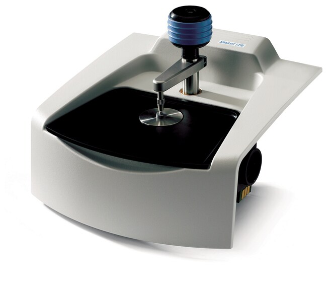
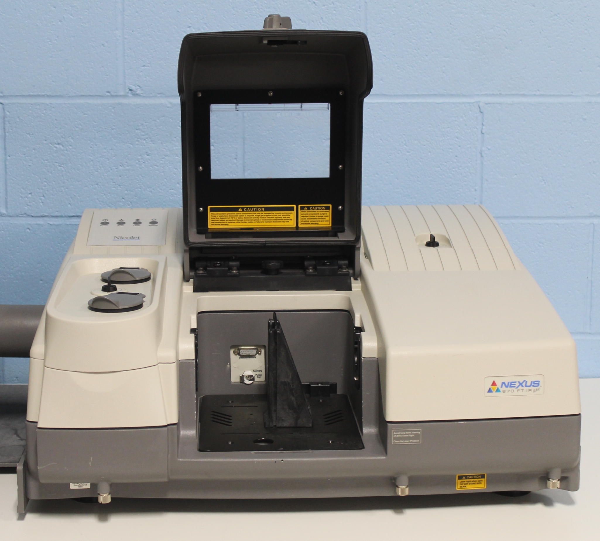
The laboratory is equipped with FT-IR, UV-Vis and FT-Raman spectrometers. The laboratory equipment and the qualifications of its staff allow the following studies to be carried out:
- determination of the structure of chemical compounds,
- spectral analysis, identification and purity control of compounds,
- low-temperature matrix-isolation spectroscopy
Below are the some examples of the extremely fast reactions where each of involved intermediates is successfully identified.
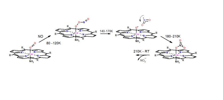
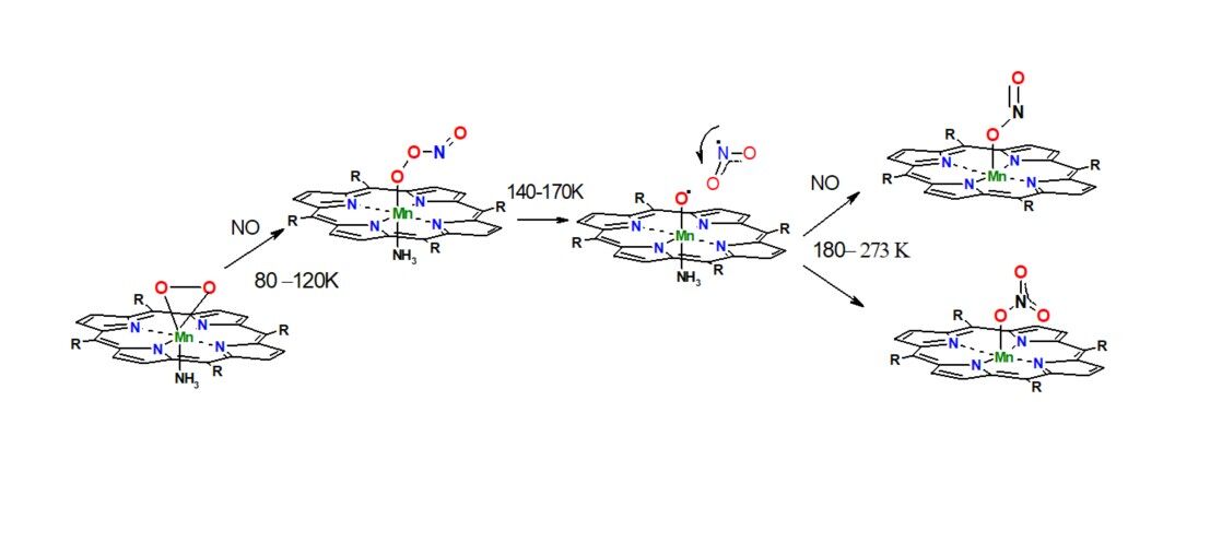
1․ In Situ FT-IR and UV−vis Spectroscopy of the Low-Temperature NO Disproportionation Mediated by Solid State Manganese(II) Porphyrinates
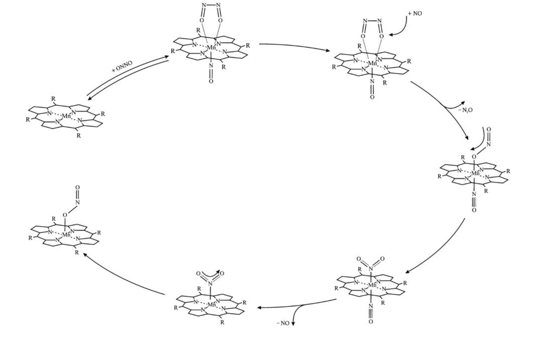
Garik G Martirosyan, Arsen S Azizyan, Tigran S Kurtikyan, Peter C Ford, Inorg. Chem., 2006, 45 (10), 4079-4087, doi.org/10.1021/ic051824q
Kurtikyan T.S. et. all., J. Amer. Chem. Soc. 2012, 134, 13861-13870.
Kurtikyan T.S. et. all., Inorg. Chem., 2013, 52, 20, 12046–12056
3. Nitric Oxide Dioxygenation by O2 Adducts of Manganese Porphyrins
Kurtikyan T.S. et. all., Inorg.Chem. 2020, 59, 17224-17233
4. Reduction of iron porphyrin nitrate to the iron nitrosyl by H2S/thiol.
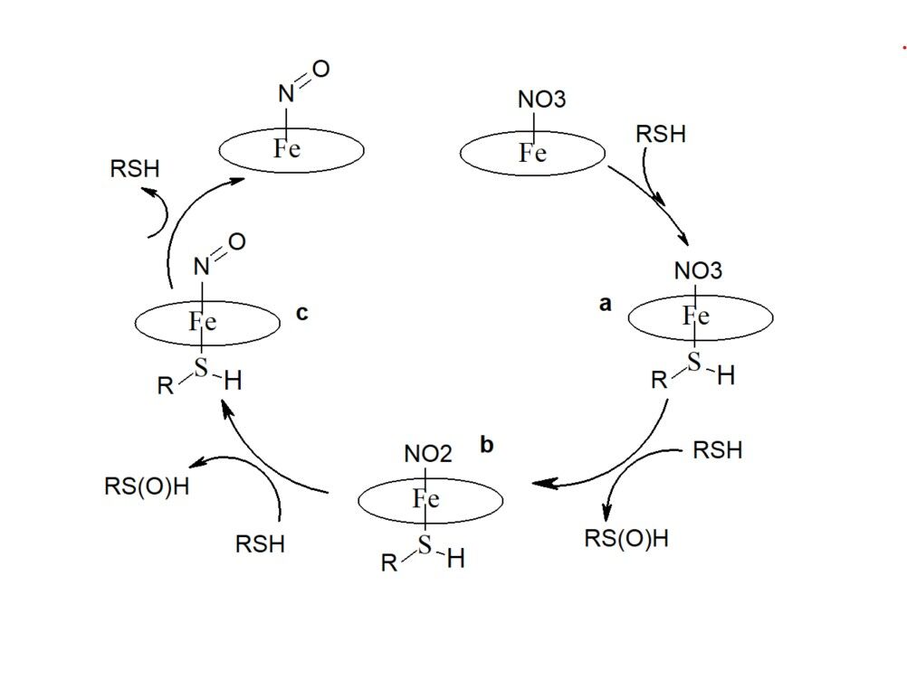 G. Martirosyan, A. A. Hovhannisyan, A. V. Iretskii, P. C. Ford., Chem. Comm., 2025, 61, 1419-1422, doi.org/10.1039/D4CC06229A,
Since 1924 one of our research directions is the study of works of art using vibrational spectroscopy methods: Raman spectroscopy and FTIR microscopy. We are cooperating with the Martiros Saryan House Museum. We are studying the pigments and binding materials used by Saryan in his paintings, trying to understand the depth and nature of the aging processes that took place in the paintings. By employing Raman spectroscopy and micro-ATR-FTIR spectroscopic imaging , successful characterization of pigments was achieved․ The results of our research reveal the presence of various pigments, including ultramarine blue, cobalt blue, cobalt cerulean blue, viridian, emerald green, cobalt green, celadonite green, cadmium yellow, chrome yellow, Venetian red, yellow ochre, red ochre, lead white, zinc white, and calcium carbonate. The findings contribute to a better understanding of Sarian’s artistic technique and provide valuable insights for the conservation and restoration of his artworks.
LUMOS II FT-IR MICROSCOPE
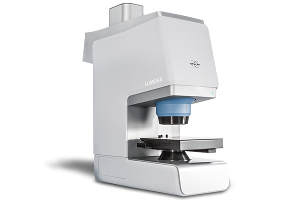
The LUMOS II FT IR Microscope from Bruker represents a significant leap forward in infrared microscopy and chemical imaging. Engineered for precision, speed, and ease of use, it delivers powerful capabilities across a wide spectrum of scientific and industrial applications.
The LUMOS II is a stand alone FT IR microscope, integrating the interferometer directly into the microscope structure. It operates seamlessly in transmission, reflection, and ATR (attenuated total reflectance) modes.In its base configuration, it features a TE MCT detector with active cooling—no liquid nitroge needed.
Users can upgrade to FPA imaging, enabling ultra fast spectral collection across 1024 pixels per scan and exceeding 50,000 spectra per minute.Vision-level spatial resolution reaches around 1.25 µm per pixel in ATR imaging.
The LUMOS II addresses a broad range of domains:
- Failure Analysis & Quality Control: Detects coatings, contaminants, polymer composition, and material defects.
- Particle & Microplastic Analysis: Combined with clustering tools, it automatically detects and classifies small particles based on chemical signature.
- Pharmaceuticals & Life Sciences: Maps APIs, excipients, and tablet cross-sections; tissue imaging becomes feasible with the ILIM variant.
- Polymers & Materials Research: Ideal for analyzing multilayer films, composites, coatings, and plastics.
- Environmental & Forensics: Reliable for trace evidence, microcontaminants, and material recognition in complex samples.
SENTERRA II RAMAN MICROSCOPES
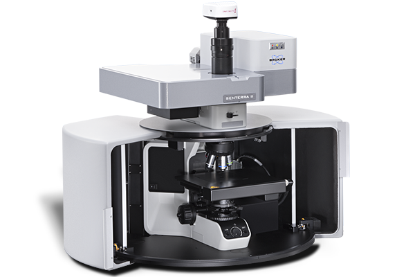
The SENTERRA II by Bruker is a cutting-edge confocal Raman microscope designed for seamless, non-stop operation, making it ideal for both industrial quality control and high-end scientific research
The versatility of SENTERRA II makes it valuable in a broad spectrum of scientific domains:
- Pharmaceuticals: Differentiates polymorphs, maps APIs/excipients, supports tablet QC.
- Material Science: Analyzes advanced materials like graphene, evaluates stress in crystalline silicon, and studies polymers and battery materials .
- Polymers & Plastics: Identifies blends, fillers, and additives, and inspects multilayer structures .
- Forensics & Art Conservation: Identifies pigments, binders, and polymers in trace evidence and fine artworks; scalable “open architecture” version enables large-area analysis.
- Environmental Science: Efficiently analyzes microplastics with automated particle detection.
•Biological Sciences: With an inverted configuration, it enables in situ Raman of live cells and tissues.
List of the important articles
Inorg. Chem., 2025, 64, 741–750, doi.org/10.1021/acs.inorgchem.4c02733
Chem. Comm., 2025, 61, 1419-1422, doi.org /10.1039/D4CC06229A,
Inorg. Chim. Acta, 2024, 566, 122031, doi.org/10.1016/j.ica.2024.122031
Macroheterocycles, 2021, 14 (4), 280-285, doi.org/10.6060/mhc214035m
Inorg. Chim. Acta, 2021, 524, 120439, doi.org/10.1016/j.ica.2021.120439
Inorg. Chem. 2020, 59, 17224-17233. doi.org /10.1021/acs.inorgchem.0c02464
Macroheterocycles, 2020, 13, 311-467. doi.org /10.6060/mhc200814k
Inorg. Chim. Acta, 2019, 495, 119011, doi.org/10.1016/j.ica.2019.119011
Inorg. Chim. Acta, 2018, 482, 894-899. doi.org/10.1016/j.ica.2018.07.044
Inorg. Chem., 2018, 57, 4795-4798, doi.org/10.1021/acs.inorgchem.8b00253
Inorg. Chem., 2016, 55, 9517-9520, doi.org /10.1021/acs.inorgchem.6b01744
Inorg. Chem., 2014, 53, 11948-11959, doi.org/10.1021/ic5014329
Inorg. Chem., 2013, 52, 12046-12056, doi.org/10.1021/ic4018689
Inorg. Chem., 2013, 52, 5201-5205, doi.org/10.1021/ic400102qGG
Inorg Biochemistry. 2013, 121, 129-133, doi.org/10.1016/j.jinorgbio.2012.12.017
Am. Chem. Soc. 2012, 134 (33), 13861-13870, doi.org/10.1021/ja305774v
Chem. Comm. 2012, 48 (99), 12088-12090, doi.org/10.1039/C2CC37337H
Laboratory devices